TAYLOR VETS
Ultrasound
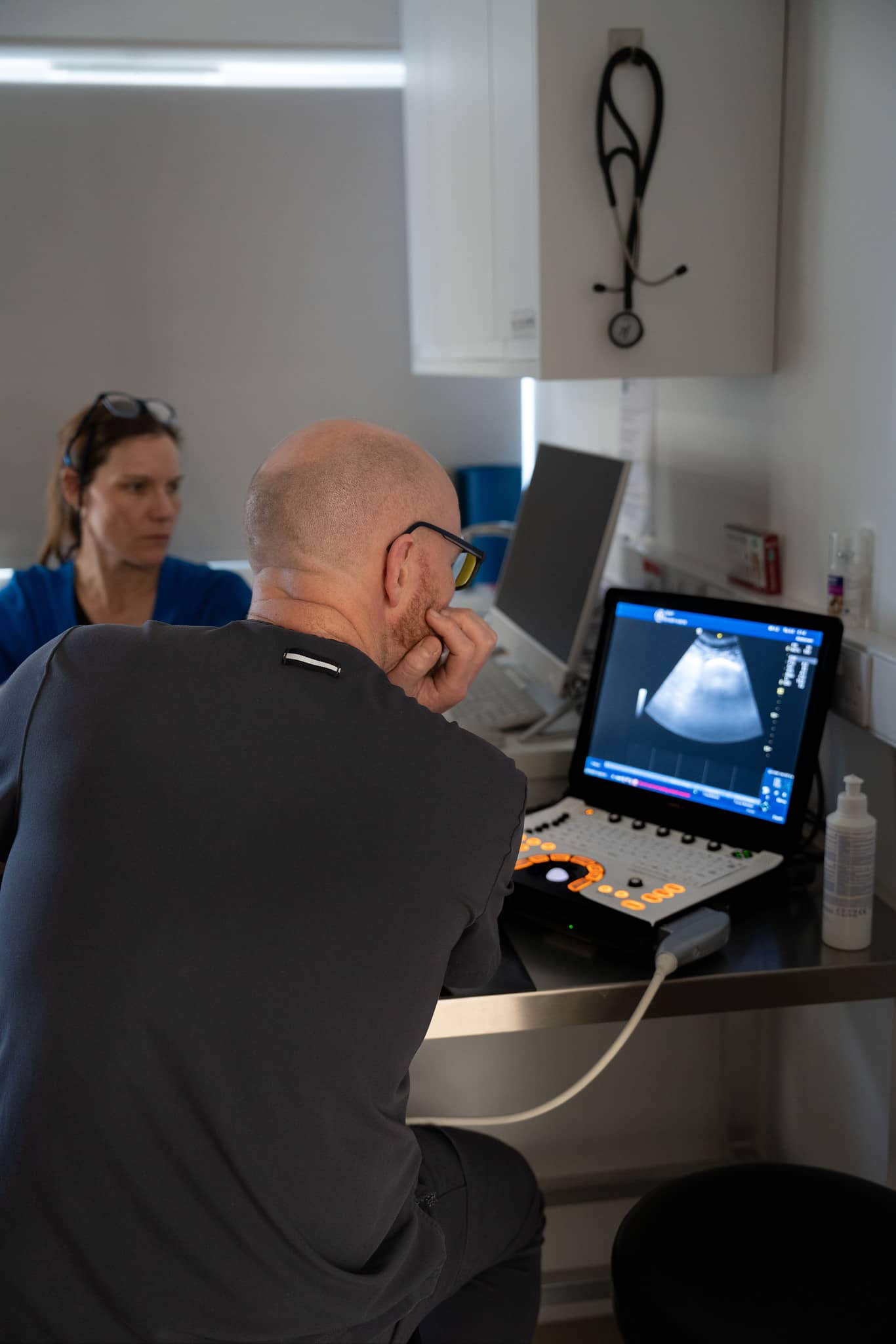
We’re proud to offer in-house ultrasound, providing clear, real-time images of soft tissue organs. This technology is perfect for examining the chest and abdominal cavities.

Primary Care Service
Ultrasound
Ultrasound procedures for cats and dogs are a safe, non-invasive, and pain-free way to examine internal organs without the use of drugs. The process uses high-frequency sound waves, which create echoes when they bounce off tissues inside the body. These echoes are then converted into electrical signals and processed by the ultrasound machine, generating a real-time image of the internal organs. This technique allows for a clearer, more detailed view of soft tissues compared to X-rays. Through ultrasound, veterinarians can assess the structure, texture, and blood flow of organs, aiding in accurate diagnosis and treatment planning.
Key Uses
- Abdominal Scans: Ultrasound is commonly used to assess the organs in the abdomen, such as the liver, kidneys, spleen, and intestines. It helps detect tumors, cysts, inflammation, and abnormal fluid accumulation.
- Cardiac Evaluations (Echocardiography): This technique allows for the examination of the heart’s structure and function. It is essential in diagnosing heart disease and monitoring heart health in animals with suspected cardiac conditions.
- Reproductive Health: Ultrasound is used to monitor pregnancies, check the health of fetuses, and investigate reproductive issues such as pyometra (uterine infection) or ovarian cysts.
- Guided Biopsies: Ultrasound is often used to guide needle biopsies for sampling tissue or fluid from abnormal areas, providing minimally invasive means of diagnosis.
- Thoracic Imaging: In some cases, it can evaluate fluid in the chest cavity or tumors in the lungs, though ultrasound is less effective in imaging through air-filled lungs compared to other modalities.
Ultrasound is an essential, versatile tool in veterinary diagnostics that can significantly aid in early diagnosis and treatment of a wide range of health issues in small animals.

Procedure Overview
The procedure is non-invasive and painless, typically requiring no sedation unless the animal is particularly anxious or in pain. The pet’s fur may be shaved in the area to allow for better contact between the ultrasound probe and the skin. The veterinarian or a trained veterinary technician will apply a conductive gel and gently glide the probe over the skin to capture real-time images of the internal organs.
Benefits:
- No Radiation Exposure: Unlike X-rays, ultrasounds do not expose pets to radiation, making it a safer option for repeated use.
- Detailed Soft Tissue Imaging: Ultrasound provides superior imaging of soft tissues, enabling better diagnosis of conditions affecting the liver, kidneys, heart, and other organs.
- Real-Time Results: The images are produced in real-time, allowing veterinarians to make immediate assessments and decisions during the procedure.